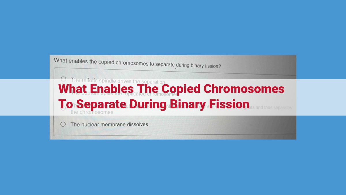
During binary fission, the copied chromosomes separate due to the coordinated action of several cellular components. The centromere, a specialized region on each chromosome, provides the attachment site for spindle fibers. These fibers, composed of microtubules, form a guiding track for the movement of chromosomes. Kinesin motor proteins transport chromosomes along the fibers towards opposite poles of the cell, while dynein motor proteins counterbalance this movement by pulling them towards the midzone. The disassembly of microtubules at the spindle poles generates the driving force for fiber movement, ultimately enabling the complete separation of copied chromosomes.
The Centromere: The Anchor for Chromosome Segregation
In the intricate tapestry of cell division, the centromere emerges as a pivotal anchor for the precise segregation of chromosomes. This specialized region of the chromosome serves as the exclusive attachment site for spindle fibers, the cellular machinery responsible for guiding chromosomes during cell division.
The centromere, a highly conserved chromosomal region, comprises a complex protein structure known as the kinetochore. This intricate assembly provides a firm foundation for the attachment of spindle fibers. Once attached, these fibers exert a pulling force that separates sister chromatids, the identical copies of each chromosome created during DNA replication.
Significance of the Centromere for Accurate Cell Division
The centromere plays a critical role in ensuring the faithful segregation of chromosomes during cell division. In mitosis, the process of cell division that produces two genetically identical daughter cells, the centromere ensures that each daughter cell receives an equal complement of chromosomes.
When centromere function is compromised, chromosome segregation errors can occur, leading to aneuploidy, a condition in which a cell possesses an abnormal number of chromosomes. Aneuploidy has been linked to a wide range of developmental disorders and diseases, including cancer.
Spindle Fibers: The Guiding Tracks for Chromosome Segregation
Within the intricate dance of cell division, spindle fibers emerge as the guiding tracks that orchestrate the separation of chromosomes. These dynamic structures, constructed from microtubules, act as the stage upon which chromosomes perform their critical journey to ensure genetic stability and cellular integrity.
Architecture and Dynamics of Spindle Fibers
Spindle fibers, also known as mitotic spindles, are polar structures that form during cell division. They consist of overlapping and interdigitating microtubules, which are long, hollow protein cylinders that provide structural support. The spindles are bipolar, meaning they have two poles, each anchored at a centrosome in the cytoplasm.
As cells prepare for division, the spindle fibers undergo remarkable changes in their architecture and dynamics. They grow, shrink, and reorganize their microtubules to form the appropriate shape and orientation for segregating chromosomes. This dynamism is essential for ensuring that each daughter cell receives the correct complement of genetic material.
Kinesin and Dynein: Motor Proteins for Chromosome Transport
Along the spindle fibers, motor proteins called kinesin and dynein play a crucial role in transporting chromosomes. Kinesin and dynein are molecular machines that use the energy from ATP hydrolysis to walk along microtubules, carrying chromosomes as their cargo.
Kinesin, a plus-end directed motor, moves towards the positive end of microtubules, while dynein, a minus-end directed motor, moves towards the negative end. This coordinated movement of chromosomes by kinesin and dynein ensures their accurate segregation to opposite poles of the cell.
Microtubule Disassembly: Driving Force for Fiber Movement
In addition to motor protein-driven movement, the disassembly of microtubules also contributes to the dynamics and force generation of spindle fibers. Microtubules are continuously assembled and disassembled, creating a treadmilling effect. The disassembly of microtubules at the spindle poles provides a pushing force that drives the separation of chromosomes.
As microtubules shorten, the chromosomes attached to them are pulled towards the poles. This disassembly-mediated force is essential for the precise segregation of chromosomes and the prevention of chromosome misalignment, which can lead to genetic instability and cell death.
Kinesin Motor Proteins: The Powerhouse of Chromosome Segregation
In the intricate world of cell division, where precision is paramount, kinesin motor proteins emerge as the unsung heroes. These molecular machines play a pivotal role in guiding the segregation of chromosomes, ensuring the faithful distribution of genetic material to daughter cells.
Structure and Function of Kinesin
Kinesins belong to a family of motor proteins that can convert chemical energy into mechanical force. They are characterized by their elongated, two-headed structure. Each head consists of a motor domain that binds to the surface of microtubules, the structural components of spindle fibers.
Contribution to Spindle Fiber Dynamics
During cell division, spindle fibers form a complex network that orchestrates chromosome movement. Kinesins interact with these fibers, contributing to their assembly, disassembly, and sliding dynamics. By moving along microtubules in a direction away from the spindle poles, kinesins generate the force that drives chromosome separation.
Implications for Chromosome Segregation
The proper functioning of kinesin motor proteins is crucial for accurate chromosome segregation. Defects in kinesin activity can lead to chromosomal misalignment and the formation of aneuploid daughter cells, which can have severe consequences for cell viability and development.
In summary, kinesin motor proteins are indispensable molecular machines that power chromosome segregation, ensuring the fidelity of cell division and the preservation of genetic information.
Dynein Motor Proteins: The Counterbalance that Ensures Chromosomal Harmony
In the intricate ballet of cell division, dynein motor proteins serve as the enigmatic counterbalance to their kin, the kinesins. These dynamic molecular machines, with their distinctive minus-end directed movement, play a pivotal role in shaping the spindle fiber dynamics that orchestrate the precise separation of chromosomes.
Unique Properties and Mechanism of Action
Dynein stands apart from its kinesin counterpart in both its structure and mechanism. Composed of multiple heavy chains adorned with various accessory subunits, dynein forms a bulky, ring-like structure. Unlike kinesin, which glides along microtubules using ATP hydrolysis, dynein employs a unique hand-over-hand mechanism. This complex molecular dance involves the sequential binding and release of dynein’s feet to adjacent tubulin subunits, propelling it towards the microtubule’s negative end.
Role in Spindle Fiber Dynamics
Within the bustling spindle fiber apparatus, dynein performs a dual role. First, it counteracts the poleward force exerted by kinesin, preventing premature separation of sister chromatids. This opposing action ensures that the chromosomes remain aligned at the metaphase plate. Second, dynein participates in microtubule disassembly, a crucial process for the disassembly of the spindle fibers once chromosome segregation is complete.
Opposing Action to Kinesin in Chromosome Separation
The interplay between dynein and kinesin is a delicate dance, essential for the equitable distribution of chromosomes to daughter cells. While kinesin propels chromosomes towards the spindle poles, dynein exerts a pulling force in the opposite direction. This tug-of-war ensures that chromosomes segregate to opposite poles, preventing chromosomal imbalances.
In conclusion, dynein motor proteins are the unsung heroes of cell division, ensuring the accuracy and fidelity of chromosome segregation. Their unique properties and opposing action to kinesin guarantee that each daughter cell receives a complete and balanced set of genetic material, paving the way for the continuation of life.
Microtubule Disassembly: The Architect of Chromosome Separation
At the heart of cell division lies a remarkable process called microtubule disassembly, a meticulously orchestrated dismantling of the cellular scaffolding that ensures the precise segregation of chromosomes. This intricate choreography is essential for maintaining the genetic integrity of dividing cells and preventing chromosomal aberrations.
The regulation of microtubule disassembly is a finely tuned dance between various cellular factors. Ubiquitin-mediated proteolysis takes center stage, targeting specific microtubule-associated proteins for degradation. This targeted destruction weakens the microtubule structure, allowing it to be broken down into its constituent tubulin subunits.
The disassembly process gives rise to a dynamic instability within the microtubule network. Shorter microtubules, with their increased susceptibility to depolymerization, are preferentially broken down, while longer, stable microtubules, attached to chromosomes at the kinetochore, are preserved. This selective disassembly generates a pulling force that physically separates the sister chromatids.
Microtubule disassembly not only facilitates chromosome separation but also contributes to the overall efficiency and fidelity of cell division. By removing excess microtubules, the cell prevents entanglements and ensures the smooth progression of chromosomal segregation. This precise dismantling process ensures that each daughter cell receives its complete complement of chromosomes, safeguarding the genetic information essential for cellular function.
Cytokinesis: The Final Cut
With the chromosomes now segregated and lined up at opposite poles of the cell, the stage is set for the final act of cell division: cytokinesis. As the name suggests, this crucial process ensures that the two new cells resulting from the division receive their fair share of cellular components.
Cytokinesis begins with the formation of a cleavage furrow, a shallow indentation that gradually deepens along the equator of the cell. This furrow is driven by the assembly and constriction of actin filaments, along with associated proteins, forming a contractile ring. As the ring tightens, it pinches the cell membrane inward, squeezing the cytoplasm into two distinct halves.
Simultaneously, microtubules extend from the opposite poles of the cell and meet at the center like a zipper. These microtubules serve as _tracks for membrane vesicles_ that contain cell wall components. As the vesicles fuse, they deposit their building blocks along the line of cleavage, gradually erecting a new cell wall that divides the two daughter cells.
The cleavage furrow, now at its deepest, eventually _severs the plasma membrane_ and cell wall, completing the _physical separation_ of the two new cells. This final cut ensures that each daughter cell inherits a complete set of chromosomes and its own independent cytoplasm and cellular components.
Cytokinesis is intimately connected to chromosome separation. The correct segregation of chromosomes during mitosis is essential for the survival of the daughter cells. If cytokinesis occurs prematurely, before the chromosomes have had a chance to separate, the resulting cells will be aneuploid, meaning they will have an abnormal number of chromosomes. This can have serious consequences for the cells’ function and viability.
