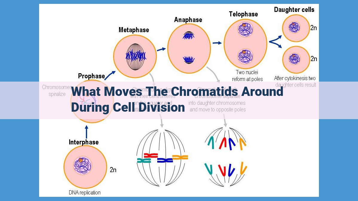
During cell division, chromatids are moved towards opposite poles by spindle fibers, which attach to the kinetochores that bind to the centromeres on the chromatids. Motor proteins like dynein and kinesin use energy from ATP to move along the microtubules, generating a pulling force that separates the chromatids. This intricate machinery ensures the precise distribution of genetic material to daughter cells.
- Importance of cell division and the need for accurate separation of chromatids
- Overview of the cell division machinery responsible for chromosome movement
Accurate Chromosome Separation: The Vital Machinery of Cell Division
In the bustling metropolis of a living cell, cell division stands as a crucial checkpoint. It ensures the precise duplication and equitable distribution of genetic material to daughter cells. At the heart of this process lies the delicate dance of specialized structures working in harmony to orchestrate the accurate separation of chromatids, the thread-like copies of chromosomes.
Imagine a team of expert movers carefully transporting precious artifacts from one mansion to another. In this analogy, the precious artifacts are chromosomes, and the team of movers represents the intricate machinery responsible for chromosome movement. This sophisticated ensemble comprises centromeres, kinetochores, microtubules, spindle fibers, and motor proteins.
Centromeres: The linchpins of chromosome segregation, centromeres are the specific locations where each chromosome is clasped by a specialized structure called a kinetochore. Think of each kinetochore as a grappling hook that firmly attaches to microtubules, the cellular highways that guide chromosome movement.
Kinetochores: These protein complexes act as the critical interface between centromeres and microtubules, ensuring the proper alignment of chromosomes during cell division. They play the role of meticulous traffic controllers, directing chromosomes to the appropriate positions within the dividing cell.
Microtubules: The scaffolding of the chromosome movement machinery, microtubules are long, slender structures that form the framework of spindle fibers. These spindle fibers extend from opposite poles of the cell, acting as tracks along which chromosomes travel.
Spindle Fibers: These bundles of microtubules are the driving force behind chromosome movement. They reach out and attach to kinetochores, pulling chromosomes towards the opposite poles of the cell. It’s like a microscopic tug-of-war, with each spindle fiber vying to claim a chromosome.
Motor Proteins: The final piece of this intricate puzzle, motor proteins are the molecular engines that power chromosome movement along microtubules. Dynein and kinesin are the two key players in this molecular symphony, ensuring that chromosomes reach their designated destinations.
Centromeres:
- Definition and role in attaching kinetochores
- Importance for proper chromosome alignment
Centromeres: The Guiding Lights of Chromosome Segregation
In the intricate dance of cell division, each chromosome must be precisely separated and distributed to the daughter cells. This critical task falls upon a remarkable structure called the centromere, the genetic epicenter of chromosome movement.
Unveiling the Centromere
The centromere is a specialized region located near the center of chromosomes. It serves as the crucial attachment point for kinetochores, protein complexes that play a vital role in chromosome segregation. Kinetochores act as molecular bridges, connecting the centromere to the microtubule spindle, the cellular machinery responsible for chromosome movement.
The Importance of Proper Alignment
The precise localization of centromeres is essential for the accurate alignment of chromosomes during division. Imagine a ballet, where each chromosome is a dancer. The centromeres are the anchors that keep them in their designated positions, ensuring they are properly oriented toward the spindle poles. When centromeres are misplaced, chromosomes can misalign, leading to genetic abnormalities and developmental disorders.
Maintaining Genomic Integrity
The centromere’s role in chromosome alignment is crucial for safeguarding the integrity of our genetic material. Faulty centromere function can result in aneuploidy, a condition where cells have an abnormal number of chromosomes. Aneuploidy is associated with a range of genetic disorders, including Down syndrome and cancer.
Future Directions
Continued research on centromere structure and function promises to deepen our understanding of cell division and its relevance to human health. By uncovering the secrets of these genetic guiding lights, we may gain insights into the causes and potential treatments for chromosome segregation errors.
Kinetochores: The Orchestrators of Chromosome Movement
In the intricate world of cell division, where life’s blueprint is duplicated and passed on, a remarkable machinery ensures that each cell receives an exact copy of the genetic material. At the heart of this intricate process lies a tiny structure known as the kinetochore, a protein complex that plays a pivotal role in guiding chromosomes to their rightful destination.
Imagine a microscopic bridge connecting the chromosomes to a network of cellular highways known as microtubules. Kinetochores serve as the anchors that tether chromosomes to these highways, ensuring their safe journey to противоположные ends of the dividing cell.
These protein complexes are not mere passive bystanders; they actively participate in the precise positioning and segregation of chromosomes. Kinetochores monitor the attachment of microtubules from opposing poles of the cell, ensuring that each chromosome is attached to microtubule fibers pulling in opposite directions. This delicate balance creates the force that aligns the chromosomes along the midline of the cell and ultimately separates them during cell division.
The kinetochore is a master of molecular choreography, orchestrating the precise movements of chromosomes. It acts as a checkpoint, preventing the cell from prematurely dividing until all chromosomes are properly attached to microtubules. This quality control mechanism safeguards against the loss or misplacement of genetic material, ensuring the integrity of the daughter cells.
In summary, kinetochores are indispensable gatekeepers of cell division, ensuring the faithful transmission of genetic information. Their ability to orchestrate chromosome movement with precision and efficiency is a testament to the remarkable complexity and elegance of life’s fundamental processes.
Microtubules: The Backbone of Chromosome Segregation
In the intricate world of cell division, where the precise separation of genetic material determines the destiny of future cells, a sophisticated machinery stands as the guardian of accuracy. Among this molecular orchestra, microtubules play a vital role, akin to the skeletal framework of a cell, ensuring the orderly movement of chromosomes.
Microtubules, delicate cylindrical structures composed of tubulin proteins, are the building blocks of the spindle fibers that orchestrate chromosome segregation during cell division. These fibers, extending from opposite poles of the cell like mitotic marionette strings, attach to the kinetochores of chromosomes—the docking stations that connect DNA to the spindle apparatus.
The formation of spindle fibers is a finely tuned ballet of molecular interactions. Microtubules undergo a process called dynamic instability—they continuously grow and shrink in length, probing their surroundings for stable connections. As they explore, they encounter kinetochores, providing a bridge between DNA and the spindle apparatus.
With microtubules firmly attached to kinetochores, the stage is set for chromosome movement. Motor proteins, the workhorses of the cell, stride along microtubules, utilizing the energy from ATP to transport chromosomes. Dynein, a motor protein that moves towards the minus end of microtubules, pulls chromosomes towards the spindle poles, while kinesin, moving towards the plus end, ensures proper alignment of chromosomes at the metaphase plate—the equatorial plane of the cell.
This intricate interplay of microtubules, kinetochores, and motor proteins ensures the precise segregation of chromosomes during cell division, safeguarding the integrity of genetic material from generation to generation.
Spindle Fibers: The Guiding Force of Chromosome Movement
In the intricate dance of cell division, meticulously orchestrated movements guide chromosomes to their designated destinations. Spindle fibers emerge as the crucial architects of chromosome segregation, the precise separation of genetic material into daughter cells.
Spindle fibers are bundles of microtubules, protein filaments that play a pivotal role in cellular structure and dynamics. During cell division, microtubules radiate from opposite poles of the cell, forming a spindle-shaped structure that serves as a scaffold for chromosome movement.
Microtubule Motor Proteins: The Movers and Shakers
Microtubules are not mere passive bystanders; they are animated by motor proteins. Dynein and kinesin, two key motor proteins, propel chromosomes along microtubule tracks. These molecular motors utilize chemical energy to generate force, enabling chromosomes to glide towards the opposite spindle poles.
The Dynamic Dance of Chromosome Segregation
With motor proteins at the helm, chromosomes embark on a carefully choreographed journey. Kinetochores, protein complexes that connect chromosomes to microtubules, act as docking stations for the motor proteins. Kinesin and dynein engage with kinetochores, pulling chromosomes towards their respective poles.
Maintaining Precision: Checkpoint Controls
To ensure faithful chromosome segregation, cells have evolved intricate checkpoint mechanisms. These safeguards prevent the cell from dividing until all chromosomes are properly aligned and attached to spindle fibers. If an error occurs, the cell delays division until the problem is resolved, minimizing the risk of genetic abnormalities.
Consequences of Spindle Fiber Dysfunction
The precise coordination of spindle fibers, motor proteins, and checkpoint mechanisms is paramount for accurate chromosome segregation. Disruptions to these processes can lead to improper chromosome distribution, resulting in genetic instability and potential developmental abnormalities. Therefore, understanding the intricacies of spindle fiber dynamics is crucial for unraveling the complexities of cell division and for devising strategies to treat diseases associated with chromosomal errors.
Motor Proteins: The Powerhouses of Chromosome Movement
Motor proteins are the unsung heroes of cell division, playing a crucial role in ensuring the precise separation of chromosomes during this critical process. These molecular motors are specialized proteins that possess the remarkable ability to move along microtubules, the structural components of the cell’s division machinery.
Among the most important motor proteins involved in chromosome movement are dynein and kinesin. Dynein is a minus-end-directed motor, meaning it moves towards the negative end of microtubules, while kinesin is a plus-end-directed motor, traveling towards the positive end.
The coordinated action of dynein and kinesin is essential for chromosome segregation. During cell division, dynein pulls chromosomes towards the spindle poles, while kinesin moves chromosomes along the spindle fibers, ensuring their proper alignment and separation.
Dynein is also involved in chromosome congression, the process by which chromosomes are aligned at the equator of the spindle. Kinesin, on the other hand, is responsible for chromosome disjunction, the separation of sister chromatids into distinct daughter cells.
These motor proteins are powered by the cellular fuel, ATP. As they hydrolyze ATP, they undergo conformational changes that drive their movement along microtubules. This ATP-dependent motility is essential for the precise and efficient movement of chromosomes during cell division.
Without the coordinated action of motor proteins, cell division would be chaotic, leading to chromosome mis-segregation and potentially disastrous consequences for the cell. These remarkable proteins are a testament to the intricate and highly organized machinery that ensures the faithful transmission of genetic material during cell division.
