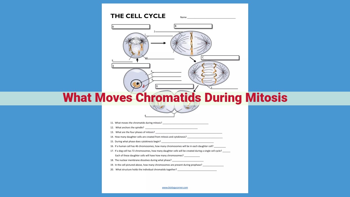
During mitosis, chromatids are moved by a complex interplay of microtubules, motor proteins, and kinetochores. Microtubules form the mitotic spindle apparatus, which connects to the kinetochores on the chromatids. Motor proteins, such as kinesin and cytoplasmic dynein, utilize energy from ATP to move chromatids along the microtubules. Kinesin pulls chromatids towards the spindle poles, while dynein separates the spindle poles and helps pull the chromatids apart. The coordinated action of these components ensures accurate segregation of genetic material during cell division.
Chromatid Movement During Mitosis: A Story of Cellular Precision
As living organisms, we embark on a continuous cycle of growth and renewal. At the heart of this process lies mitosis, a remarkable dance of cellular division that ensures the faithful transmission of genetic material to daughter cells. Within this intricate ballet, the precise movement of chromatids plays a pivotal role.
Chromatids: Carriers of Genetic Heritage
Imagine chromatids as tiny threads of DNA, carrying the blueprint for our genetic makeup. During mitosis, these threads undergo an elaborate journey, meticulously guided by a symphony of cellular machinery. The proper execution of this movement is paramount for the integrity of our genetic inheritance and the very survival of life itself.
Kinetochore: The Orchestrator of Chromatid Movement during Mitosis
In the intricate dance of mitosis, the division of a cell into two identical daughter cells, the precise movement of chromatids is crucial. And at the heart of this movement lies a tiny structure called the kinetochore.
The kinetochore is a protein complex that attaches spindle fibers to each sister chromatid, providing a physical bridge for their separation during mitosis. These spindle fibers are microtubules, long, hollow tubes that form the framework of the spindle apparatus, a structure that guides chromosome movement.
The kinetochore acts as a molecular gatekeeper, ensuring that each chromatid is properly attached to microtubules before the chromosomes begin their journey to opposite poles of the cell. This attachment occurs at a specific region of the chromatid known as the centromere.
The interaction between kinetochores and microtubules is a dynamic process regulated by a range of molecular players. This interaction ensures that only properly attached chromatids are pulled apart, preventing errors that could lead to genetic abnormalities and cell malfunctions.
The kinetochore is a critical component of mitosis, ensuring the equal distribution of genetic material to daughter cells. Its role in coordinating the movement of chromosomes is essential for maintaining the integrity of our genetic code and the health of our cells.
Microtubules:
- Structure and composition of microtubules
- Formation of the spindle apparatus and its role in chromatid movement
Microtubules: The Architectural Framework for Chromatid Movement in Mitosis
In the intricate dance of mitosis, where cells divide and replicate their genetic blueprint, microtubules play a starring role. These dynamic, hollow structures are the building blocks of the spindle apparatus, the scaffolding that orchestrates the precise movement of chromatids.
Microtubules, composed of the protein tubulin, are tube-like structures that can assemble and disassemble rapidly. During mitosis, they polymerize in a meticulous manner, forming the spindle apparatus. This spindle, shaped like an hourglass or two poles connected by a central spindle, provides the tracks for chromatid movement.
The structure of microtubules is crucial for their function. They consist of alternating subunits of alpha and beta tubulin, arranged in a helical pattern. The polarity of microtubules, with a plus end and a minus end, determines their direction of growth and movement. During spindle formation, minus ends converge at the spindle poles, while plus ends extend toward the equator of the spindle, creating the fibrous framework that will guide chromatids.
The spindle apparatus plays a pivotal role in chromatid movement. Microtubule bundles emanating from opposing spindle poles attach to the kinetochores of sister chromatids, specialized structures at the centromere of each chromosome. This attachment ensures that each chromatid is pulled equally to opposite spindle poles, ensuring the faithful segregation of genetic material.
Chromatid Movement During Mitosis: The Orchestration of Motor Proteins
During the intricate dance of mitosis, the faithful segregation of genetic material is paramount. Chromatids, the DNA-laden arms of chromosomes, must undergo a precise journey to ensure each daughter cell receives an identical set of chromosomes. This intricate process involves the interplay of various cellular components, including motor proteins.
Motor proteins, the workhorses of the cell, are specialized proteins that navigate along microtubules, the structural highways that provide the framework for cell division. These molecular machines possess remarkable properties that enable them to transport cellular cargo, including chromatids, towards their designated destinations.
Among the motor proteins involved in chromatid movement are kinesin and dynein. Kinesins move towards the plus ends of microtubules, pulling chromatids towards the spindle poles. These poles represent the opposite ends of the mitotic spindle, an apparatus assembled from microtubules that guides chromosome movement.
On the other hand, dynein, a highly specialized motor protein, moves towards the minus ends of microtubules. Dynein is primarily responsible for separating the spindle poles during anaphase, the stage of mitosis when sister chromatids are pulled apart. Additionally, dynein aids in the initial attachment of kinetochores, the protein complexes that connect chromatids to microtubules, to the spindle apparatus.
The attachment of kinetochores to microtubules is critical for the proper segregation of chromosomes. Motor proteins, through their interaction with kinetochores, ensure that each chromatid is securely attached to a spindle fiber. This attachment provides the necessary tension to separate sister chromatids during anaphase.
The coordinated interplay of kinesin and dynein, along with other cellular components involved in chromatid movement, is essential for the precise partitioning of genetic material during mitosis. This intricate process ensures the faithful transmission of genetic information, preventing errors that could lead to developmental abnormalities and genetic disorders.
Cytoplasmic Dynein: The Powerhouse Behind Chromosome Separation
In the intricate dance of mitosis, a crucial player emerges: cytoplasmic dynein, a motor protein that orchestrates the separation of spindle poles and the precise pulling apart of chromosomes. This remarkable protein holds the key to ensuring the equitable distribution of genetic material during cell division.
Dynein’s story unfolds at the heart of the cell, within the spindle apparatus. Picture a network of microtubules, a scaffolding-like structure that serves as the stage for mitosis. Dynein molecules, acting like tiny engines, crawl along these microtubule tracks, exerting forces that orchestrate the movement of chromosomes.
As dynein binds to the spindle poles, its power is unleashed. It begins to walk towards the opposite pole, dragging the microtubule with it. This pulling force not only separates the spindle poles but also creates tension within the spindle fibers, providing the power to pull the chromosomes apart.
Dynein’s role extends beyond pole separation. It also participates in a tug-of-war with kinesin, another motor protein that moves chromosomes towards the center of the spindle. The combined action of dynein and kinesin ensures the precise alignment and segregation of chromosomes, ensuring that each daughter cell receives an identical genetic blueprint.
Cytoplasmic dynein’s unwavering dedication to mitosis makes it an indispensable player in the symphony of cell division. Its precise movements and coordination with other proteins safeguard the integrity of genetic material, providing the foundation for the growth and development of organisms.
Kinesin: The Powerhouse of Chromosome Movement
In the intricate dance of mitosis, where chromosomes divide and cells split, kinesin stands as a microscopic marvel, guiding chromosomes to their appointed poles. This motor protein plays a crucial role in the precise orchestration of cell division, ensuring the equal distribution of genetic material.
Kinesin’s structure is a testament to its specialized function. Composed of a motor domain that binds to microtubules and a stalk domain that extends like a bridge, kinesin is poised to move along microtubule tracks. Like a microscopic train, kinesin transports chromosomes, hitching them to its stalk domain and pulling them towards the spindle poles.
The energy for kinesin’s movement comes from the breakdown of ATP molecules. As ATP binds to the motor domain, it triggers a change in conformation, causing the motor domain to step forward along the microtubule. This stepping motion, repeated over and over, propels the chromosome-laden kinesin towards the spindle poles.
Kinesin’s role in mitosis is paramount. It ensures that each daughter cell receives a complete set of chromosomes, preventing genetic abnormalities and maintaining genomic stability. Without kinesin’s tireless efforts, cell division would be chaotic and potentially disastrous.
In conclusion, kinesin is a captivating example of the molecular machinery that underpins the fundamental processes of life. Its meticulous movement of chromosomes during mitosis ensures the faithful transmission of genetic information from one generation of cells to the next.
Chromatid Arms: The Guiding Lights of Chromosome Movement
In the intricate dance of mitosis, the division of a cell into two identical daughter cells, the precise movement of chromatids is pivotal. Each chromatid is a condensed form of a replicated chromosome, and its journey to its designated pole is guided by an unsung hero: the chromatid arm.
Imagine the chromatid arms as microscopic docking stations, where microtubules, the cellular highways, come to connect. These microtubules form the spindle apparatus, a complex structure that orchestrates the movement of chromatids.
The chromatid arms are strategically positioned along the chromatid, providing multiple attachment points for microtubules. These attachment sites are crucial for creating the forces necessary for chromatid separation.
The Pull and the Push
The movement of chromatids is driven by two types of motor proteins: kinesin and dynein. Kinesin, the workhorse of the system, pulls chromatids towards the spindle poles, while dynein, the master of separation, pushes the poles apart.
These motor proteins use the energy from ATP to walk along microtubules, dragging chromatids behind them. The coordinated action of kinesin and dynein ensures that chromatids are separated and transported to opposite poles of the dividing cell.
Precision in Motion
The accuracy of chromatid movement is vital for cell division. If chromatids fail to separate correctly, the resulting daughter cells will have an incorrect number of chromosomes, leading to genetic abnormalities.
The precise attachment of microtubules to chromatid arms ensures that each chromatid is captured and transported to the correct pole. This intricate mechanism guarantees the faithful transmission of genetic information to future generations.
