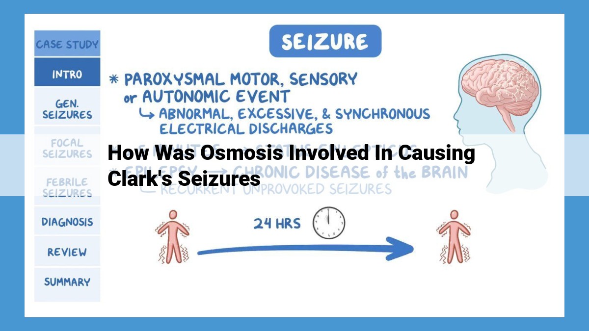
Osmosis, the movement of water across semipermeable membranes, has been implicated in Clark’s seizures. Hypertonicity, an imbalance in water potential, can cause water to flow out of brain cells, leading to crenation and reduced brain volume. This osmotic stress can disrupt neuronal excitability and lower the seizure threshold. Aquaporins, channels that facilitate water movement, and the blood-brain barrier play crucial roles in regulating osmotic balance. Further research is needed to establish a causal relationship between osmosis and seizures and explore therapeutic strategies targeting osmotic imbalances.
- Describe Clark’s seizures and their classification as epilepsy.
- State the hypothesis that osmosis played a role in causing these seizures.
Clark’s Seizures: Unraveling the Role of Osmosis
In the realm of medical mysteries, the case of Clark’s seizures stands as a testament to the enigmatic nature of the human body. Clark’s seizures, characterized by uncontrollable jerking movements and loss of consciousness, confounded medical experts for years. However, a groundbreaking hypothesis emerged, suggesting that a cellular phenomenon known as osmosis might hold the key to understanding these puzzling episodes.
Osmosis: The Dance of Water
Envision a cellular playground where water molecules, the lifeblood of our bodies, engage in a ceaseless dance. Osmosis, the driving force behind this dance, orchestrates the movement of water across semipermeable membranes that guard our cells. It’s all about balancing the scales, ensuring that the concentration of dissolved particles on both sides of the membrane remains in equilibrium.
Water’s Dance within Cells
Within our cells, water embarks on a strategic exchange, flowing in and out through specialized channels called aquaporins. This constant influx and efflux of water plays a vital role in maintaining cellular homeostasis, the delicate balance that keeps our cells thriving.
Hypertonicity and Hypotonicity: When Water’s Dance Goes Awry
But when water’s dance goes amiss, cells can face dire consequences. Hypertonicity, a condition where the concentration of dissolved particles outside the cell is higher, causes water to flee the cell in search of equilibrium. This can lead to cell shrinkage, a potentially damaging state. Conversely, hypotonicity, where the concentration of dissolved particles outside the cell is lower, encourages water to rush in, causing cell swelling that can lead to cell lysis.
**The Blood-Brain Barrier: Guardi
Understanding Osmosis
- Define osmosis and explain its mechanism of water movement.
- Explain water potential, semipermeable membranes, and the role of diffusion in osmosis.
Understanding Osmosis: The Vital Force Behind Water Movement
Imagine a cell, the building block of life, like a tiny water balloon. Its delicate membrane acts as a semipermeable barrier, allowing some substances to pass through while blocking others. This barrier plays a crucial role in a fascinating process called osmosis, the movement of water across membranes.
Osmosis is driven by the difference in water potential between two compartments. Water potential is a measure of how much water molecules want to move from one place to another, much like the pressure difference that drives air through a straw. When water potential is higher on one side of the membrane, water molecules rush from the high to the low potential side to equalize it.
This movement of water is essential for cells. Cells constantly exchange nutrients, waste products, and other substances with their surroundings, and water serves as the transportation medium. Aquaporins, tiny channels in the membrane, facilitate this water movement, allowing water molecules to pass through the membrane quickly and efficiently.
Hypertonicity and hypotonicity are two important concepts in osmosis. When the water potential is lower outside a cell compared to inside, the cell is said to be hypertonic. In such conditions, water moves out of the cell, causing it to shrink and potentially even become crenated (wrinkled). Conversely, when the water potential is higher outside the cell, the cell is hypotonic. Water rushes into the cell, causing it to swell and potentially even burst.
Osmosis plays a vital role in maintaining cell homeostasis, the delicate balance that keeps cells functioning properly. It ensures that cells have the right amount of water to carry out their essential tasks and that they are not damaged by excessive water influx or efflux.
Water Influx and Efflux in Cells: A Vital Dance for Cell Health
Water is the lifeblood of our cells, constantly flowing in and out to maintain a delicate balance that ensures our bodies function properly. This dynamic exchange, known as osmosis, plays a crucial role in regulating cell volume and homeostasis.
Aquaporins: The Gatekeepers of Water Movement
Imagine aquaporins as microscopic channels embedded within cell membranes. These specialized proteins are responsible for facilitating the movement of water molecules across the membrane, a process known as osmosis. Without aquaporins, water movement would be severely restricted, impairing cell function and potentially leading to cell damage.
Osmotic Pressure: The Force that Drives Water Flow
Osmotic pressure is the force that drives the movement of water across a semipermeable membrane. When two solutions with different concentrations of dissolved substances are separated by a semipermeable membrane, water molecules move from the solution with a lower concentration (lower osmotic pressure) to the solution with a higher concentration (higher osmotic pressure). This movement continues until the concentrations on both sides of the membrane are equal, or an equilibrium is reached.
The Importance of Water Influx and Efflux
The constant influx and efflux of water maintain cell homeostasis. When a cell takes in too much water, it swells. Conversely, when a cell loses too much water, it shrinks. This delicate balance is essential for maintaining proper cell function.
For example, in red blood cells, water influx and efflux help regulate the concentration of hemoglobin, which carries oxygen throughout the body. If red blood cells swell too much, they may burst, releasing hemoglobin into the bloodstream and causing a condition known as hemolysis. On the other hand, if red blood cells shrink too much, they may not be able to carry enough oxygen to meet the body’s needs.
Osmosis: The Potential Culprit Behind Clark’s Seizures
Hypertonicity and Hypotonicity: A Tale of Two Extremes
Within the realm of osmosis, two opposing forces emerge: hypertonicity and hypotonicity. Let’s delve into their distinct characteristics and their impact on cells.
Hypertonicity: When the Outside Packs a Punch
Imagine a cell immersed in a hypertonic environment. The high solute concentration outside the cell creates a situation where water molecules are drawn out of the cell. This water loss leads to cell shrinkage. In severe cases, the cell may even undergo crenation—a process where the cell membrane puckers and folds.
Hypotonicity: When the Cell Swells with Abundance
In contrast, a hypotonic environment boasts a low solute concentration. Water molecules swiftly rush into the cell, eager to balance the disparity. As the cell absorbs water, it swells and expands. If the water influx is excessive, the cell membrane may stretch beyond its limits, ultimately rupturing and causing lysis—the cell’s demise.
The Blood-Brain Barrier: A Guardian of the Mind
Nestled within the intricate web of our brain’s anatomy lies a vital protective layer known as the blood-brain barrier (BBB). This highly specialized shield plays a critical role in safeguarding the delicate neurons that orchestrate our thoughts, feelings, and actions.
Comprising an intricate network of cells and blood vessels, the BBB serves as a vigilant gatekeeper, regulating the entry of substances into the brain. It carefully selects molecules that are essential for neuronal function while filtering out potentially harmful compounds. This selectivity ensures that the brain is shielded from toxins, pathogens, and other threats that could disrupt its delicate balance.
The neurovascular unit, a harmonious partnership between neurons, glial cells, and blood vessels, forms the foundation of the BBB. This intricate network ensures the efficient delivery of nutrients and oxygen to the brain while maintaining its protective barrier. The choroid plexus, a specialized structure within the brain’s ventricles, produces the cerebrospinal fluid that bathes and nourishes the brain and spinal cord, further contributing to the BBB’s protective function.
The ependymal cells, lining the ventricles, collaborate with the choroid plexus to diligently monitor the composition of the cerebrospinal fluid. Acting as alert sentinels, they detect and remove any potential threats before they can infiltrate the sensitive brain tissue.
The BBB’s remarkable ability to regulate the brain’s environment is crucial for maintaining its delicate equilibrium. This protective barrier ensures that the right substances reach the brain at the right time, while harmful intruders are kept at bay. It is a testament to the body’s intricate design, safeguarding the most precious organ of all—our mind.
Neuronal Excitability and Seizures
Neurons, the fundamental units of our brain, are like tiny electrical circuits that transmit signals throughout our bodies. These signals are triggered by changes in the electrical potential across the neuron’s membrane, a process known as neuronal excitability.
Ion Channels, Neurotransmitters, and Synaptic Plasticity
Ion channels are pores in the neuron’s membrane that allow charged particles, such as sodium and potassium ions, to flow in and out of the cell. This controlled flow of ions creates the electrical potential difference that underlies neuronal excitability. Neurotransmitters are chemical messengers that bind to receptors on the neuron’s membrane, triggering the opening or closing of ion channels and altering excitability. Synaptic plasticity, the ability of synapses (the connections between neurons) to strengthen or weaken over time, also plays a crucial role in neuronal excitability.
The Relationship between Neuronal Excitability and Brain Activity
The level of neuronal excitability governs the frequency and intensity of brain activity. When excitability is high, neurons are more likely to fire and transmit signals, leading to increased brain activity. Conversely, when excitability is low, neurons are less likely to fire, resulting in decreased brain activity.
The Role of Neuronal Excitability in Seizures
Epilepsy is a neurological disorder characterized by recurrent seizures, sudden, uncontrolled bursts of electrical activity in the brain. During a seizure, neurons in the brain become hyperexcitable, firing excessively and asynchronously. This abnormal electrical activity can manifest as a variety of physical symptoms, including convulsions, muscle spasms, and loss of consciousness.
Seizure Threshold
- Define seizure threshold and its significance in epilepsy.
- Explain the role of anticonvulsants in raising seizure threshold.
- Discuss electroencephalography (EEG) as a tool for monitoring seizure activity.
Exploring the Role of Osmosis in Clark’s Seizures
Clark’s life was marked by frequent seizures, characterized as epilepsy. A groundbreaking hypothesis emerged, suggesting that osmosis, the movement of water across membranes, played a crucial role in triggering these seizures.
Understanding Osmosis: The Water’s Journey
Osmosis is the process by which water flows from areas of low solute concentration to areas of high solute concentration. Semipermeable membranes, like cell membranes, allow water molecules to pass through while blocking larger molecules. This water movement maintains the balance of water potential, a measure of water’s tendency to move.
Water in and Out of Cells: A Delicate Dance
Cells rely on the precise regulation of water movement. Aquaporins, specialized proteins, facilitate water’s entry and exit. Osmotic pressure, the force exerted by the difference in solute concentration across a membrane, governs cell volume. When external conditions are more concentrated (“hypertonic”), water leaves the cell, causing shrinkage. Conversely, when conditions are less concentrated (“hypotonic”), water enters the cell, potentially causing swelling and even bursting.
The Blood-Brain Barrier: A Protective Layer
The brain’s unique blood-brain barrier shields it from potentially harmful substances in the bloodstream. Intricate mechanisms, including tight junctions and specialized cells, maintain this protective barrier, ensuring the brain’s delicate internal environment.
Neuronal Excitability: The Spark of Thought
The brain’s neurons rely on electrical signals for communication. Ion channels, protein pores in cell membranes, allow for the exchange of ions, generating the electrical pulses that transmit information. Neurotransmitters, chemicals that facilitate communication between neurons, play a pivotal role in neuronal excitability.
Seizure Threshold: The Line Between Order and Chaos
Seizure threshold refers to the level of excitation required to trigger a seizure. Anticonvulsants, medications commonly used to treat epilepsy, work by raising the seizure threshold, making it more difficult for seizures to occur. Electroencephalography (EEG), a non-invasive technique, provides a window into brain activity, helping doctors monitor seizure patterns and assess treatment efficacy.
Osmosis and Clark’s Seizures: A Possible Connection
The hypothesis linking osmosis to Clark’s seizures postulates that hypertonicity within the brain, resulting in excessive water loss from neurons, could lead to neuronal hyperexcitability and ultimately trigger seizures. Aquaporins and the blood-brain barrier may play critical roles in this process. The interplay between neuronal excitability and osmotic changes remains a fascinating area of research, with the potential for new insights into seizure mechanisms and therapeutic interventions.
Osmosis and Clark’s Seizures: A Hypothesis
Clark, a young and brilliant artist, had a dark secret. His seizures, a relentless torment that haunted him since childhood, cast a long shadow over his otherwise vibrant life. Despite numerous medical interventions, the cause of his seizures remained an enigma. But a breakthrough emerged when researchers stumbled upon a tantalizing hypothesis: osmosis might be the hidden culprit.
Understanding Osmosis: The Movement of Water
Osmosis, a fascinating natural process, drives the movement of water across semipermeable membranes. When two solutions with different concentrations are separated by a membrane that allows water to pass through (but not dissolved particles), water flows from the area of lower solute concentration to the area of higher solute concentration. This movement balances the concentrations on both sides of the membrane.
Osmotic Pressure and Its Effects on Cells
The difference in solute concentration between two solutions creates a force called osmotic pressure. When cells are placed in a hypertonic solution (one with a higher solute concentration than inside the cell), water moves out of the cell in an attempt to equalize the concentrations. This causes the cell to shrink, a phenomenon known as crenation. Conversely, when cells are placed in a hypotonic solution (lower solute concentration outside the cell), water rushes in, causing the cell to swell and potentially burst, a process called lysis.
The Blood-Brain Barrier and Neuronal Excitability
The brain, our most precious organ, is shielded from harmful substances by the blood-brain barrier. This specialized network of cells regulates the passage of molecules between the blood and the central nervous system. As such, it plays a crucial role in maintaining a stable environment for the brain’s delicate neurons.
Seizures arise from uncontrolled neuronal activity, leading to abnormal electrical discharges in the brain. These discharges can be triggered by various factors, including changes in ion concentrations and neurotransmitter imbalances. Osmotic changes in the brain’s extracellular fluid can influence neuronal excitability, potentially contributing to seizure activity.
The Hypothesis: Osmosis and Seizures
The hypothesis that osmosis may have contributed to Clark’s seizures stems from the observation that his seizures tended to occur during periods of hypertonicity in the brain. Hypertonicity can result from dehydration, certain medications, or even hormonal changes.
In such conditions, water may be drawn out of neuronal cells, causing the cells to shrink and their membranes to become more excitable. This increased excitability lowers the seizure threshold, making the brain more prone to electrical discharges.
Aquaporins and the Role of the Blood-Brain Barrier
Aquaporins, specialized channel proteins, facilitate the movement of water across cell membranes. They play a pivotal role in osmoregulation, including in the brain. The blood-brain barrier contains aquaporins that regulate water influx and efflux in the brain’s extracellular fluid.
Disruptions in aquaporin function or alterations in the integrity of the blood-brain barrier could potentially lead to osmotic imbalances and affect neuronal excitability, increasing the likelihood of seizures.
Future Research and Implications
The hypothesis that osmosis played a role in Clark’s seizures offers a novel perspective on seizure etiology. Further research is needed to validate this hypothesis and explore its implications for the diagnosis and treatment of epilepsy. Understanding the role of osmosis in seizures could pave the way for new therapeutic strategies aimed at regulating water balance in the brain and preventing seizure activity.
