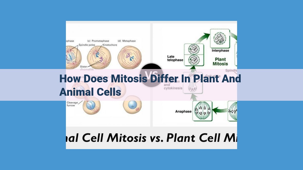
Mitosis differs in plant and animal cells primarily in cytokinesis. Plant cells form a cell plate via the phragmoplast, a microtubule-based structure unique to plants. In contrast, animal cells utilize cleavage furrow constriction. Furthermore, centrosomes, which organize spindle fibers, differ between the two cell types. While animal cells possess distinct centrosomes, plant cells lack true centrosomes and rely on spindle fibers organized from multiple sites. These variations in cytokinesis mechanisms and spindle fiber organization reflect the distinct challenges posed by the presence or absence of a cell wall during cell division.
Cell Plate Formation: Nature’s Zipper in Plant Cell Division
In the realm of cell division, where the dance of mitosis unfolds, cytokinesis emerges as the grand finale, a delicate process that divides the cytoplasm and distributes genetic material evenly between two daughter cells. For plant cells, this orchestration of cytokinesis requires a unique structure known as the cell plate.
Imagine the cell plate as a cellular zipper, gradually closing in on itself to segregate the cytoplasm. This intricate structure is formed when vesicles containing cell wall components align along the equator of the dividing cell. As these vesicles fuse, they deposit their contents to form the nascent cell wall.
Cell wall formation is a defining characteristic of plant cells during cytokinesis. Unlike animal cells, the presence of a cell wall poses a unique challenge, requiring a specialized mechanism to ensure that the daughter cells inherit their own complete cell wall.
The Phragmoplast: A Plant-Specific Cytokinesis Orchestrator
In the realm of cell division, plant cells embark on a unique journey unlike their animal counterparts. Cytokinesis, the final stage of cell division that separates the cytoplasm, presents a distinct challenge for plant cells due to their rigid cell walls. Enter the phragmoplast, a remarkable microtubule-based structure that plays a pivotal role in plant cell cytokinesis.
Imagine the phragmoplast as a cellular choreographer, guiding the formation of a new cell wall that will divide the cytoplasm and create two distinct daughter cells. This intricate structure consists of microtubules, long, thin protein filaments that form an array resembling a cell plate.
As the phragmoplast expands, it facilitates the movement of vesicles containing cell wall material towards the center of the cell. These vesicles fuse together, depositing their contents to form the new cell wall, much like bricklayers building a wall. This process ensures that the two daughter cells will have their own independent cell walls, a crucial step in maintaining cell integrity.
The phragmoplast not only facilitates cell wall formation but also plays a vital role in the separation of chromosomes. During mitosis, the chromosomes are organized on a structure called the spindle apparatus. In plant cells, the phragmoplast is closely associated with the spindle fibers, helping to ensure that the chromosomes are distributed equally between the daughter cells.
Once the phragmoplast has successfully completed its mission, it disassembles, leaving behind two new daughter cells with their own distinct cytoplasm and cell walls. The plant cell cycle can then proceed, giving rise to new cells that will contribute to the growth and development of the plant.
Centrosomes and Spindle Fibers: Key Players in Cytokinesis
In the realm of cell division, centrosomes and spindle fibers play pivotal roles in the intricate dance known as cytokinesis. These cellular structures orchestrating the meticulous division of the cytoplasm during cell division.
Centrosomes: The Microtubule Orchestrators
Imagine centrosomes as tiny organelles, the architects of cellular division. They are responsible for organizing microtubules, the structural components that form the framework of the cell. These microtubules serve as the tracks along which chromosomes travel during cell division.
Spindle Fibers: The Chromosome Separators
Spindle fibers, composed of microtubules, are the driving force behind chromosome separation. During mitosis, they attach to chromosomes and pull them apart, ensuring an equitable distribution of genetic material between daughter cells.
Differences in Plant and Animal Cells
While centrosomes and spindle fibers are essential for cytokinesis in both plant and animal cells, their organization and function differ. In animal cells, centrosomes are located at opposite poles of the cell, acting as anchors for spindle fibers. In plant cells, however, centrosomes are absent. Instead, spindle fibers are organized by a unique structure called the phragmoplast, a microtubule-based scaffold that facilitates cell plate formation.
Microtubule Assembly and Disassembly
The dynamic nature of microtubule assembly and disassembly is crucial for cytokinesis. During mitosis, microtubules polymerize and depolymerize in a coordinated manner, allowing spindle fibers to lengthen and shorten, driving chromosome movement and cell division.
Centrosomes and spindle fibers are essential components of the cellular machinery responsible for cytokinesis. Their intricate organization and function ensure the precise division of cytoplasm, a critical step in cell division and the propagation of life.
Cytokinesis in Plant and Animal Cells: Unique Challenges
Cytokinesis, the final stage of cellular division, ensures the equitable distribution of genetic material to daughter cells. However, plant and animal cells face unique challenges during cytokinesis due to their distinct cellular structures.
In animal cells, the cytoplasm divides through a process called furrow formation. A contractile ring of actin and myosin filaments pinches the cell membrane inward, eventually dividing the cytoplasm.
Plant cells, on the other hand, have an additional barrier to overcome: the rigid cell wall. To accommodate this, plant cells employ a specialized structure called the phragmoplast. The phragmoplast is a microtubule-based scaffold that guides the formation of the cell plate, a new cell wall that divides the cytoplasm.
The cell plate grows from the center of the cell outward, guided by the phragmoplast. As it expands, vesicles containing cell wall materials fuse with the growing cell plate, strengthening and completing the new cell wall.
Centrosomes and spindle fibers, organelles involved in cell division, differ between plant and animal cells. In animal cells, centrosomes are responsible for organizing microtubules, including the spindle fibers that separate chromosomes during cell division. In plant cells, centrosomes are absent, and spindle fibers are organized by the phragmoplast.
Despite these differences, the fundamental goal of cytokinesis remains the same in both plant and animal cells: to ensure the proper distribution of genetic material and the creation of two independent daughter cells.
