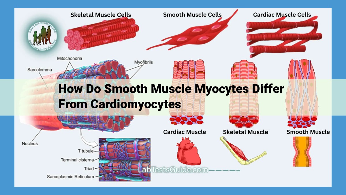
Smooth muscle myocytes differ from cardiomyocytes in morphology (spindle-shaped vs. striated), arrangement (sheets vs. intercalated discs), and contraction characteristics. Smooth myocytes have less organized actin-myosin filaments, leading to slower contractions. They lack sarcomeres and intercalated discs, enabling sustained and non-rhythmic contractions. Innervated by the autonomic nervous system, they control involuntary functions like vascular tone and motility. Unlike cardiomyocytes, smooth muscle myocytes can divide, allowing for repair and growth.
Morphology and Arrangement
- Describe the spindle-shaped morphology and sheet-like arrangement of smooth muscle myocytes.
- Contrast with the striated appearance and intercalated discs of cardiomyocytes.
Morphology and Arrangement: Unraveling the Unique Features of Smooth Muscle
In the realm of muscles, two distinct types exist: smooth muscle and cardiomyocytes. While both serve essential functions, their architectural blueprints differ markedly. Let’s delve into the morphology and arrangement that shape their distinct roles.
Spindle-Shaped Myocytes: A Flexible Framework
Smooth muscle myocytes, the building blocks of smooth muscle, possess a unique spindle-shaped morphology. These elongated cells align themselves in sheet-like formations, creating a flexible and versatile tissue. Unlike cardiomyocytes, which showcase a striated appearance due to their organized arrangement of proteins, smooth muscle myocytes lack this characteristic feature.
Contrast with Striated Cardiomyocytes
In stark contrast, cardiomyocytes showcase a highly organized appearance. They are cylindrical in shape and arranged end-to-end, forming intercalated discs. These specialized junctions facilitate synchronous contraction, ensuring the rhythmic beating of the heart. The absence of such discs in smooth muscle myocytes reflects their distinct mode of contraction.
Actin and Myosin Filaments: A Tale of Contraction in Smooth Muscle
Smooth muscle cells, the unassuming workhorses of our bodies, possess a unique set of characteristics that enable them to perform their vital functions. One key feature that distinguishes them from striated muscle cells lies in the arrangement of their actin and myosin filaments, the fundamental components responsible for muscle contraction.
Unlike the highly organized, striated pattern found in striated muscle cells, the actin and myosin filaments in smooth muscle cells are arranged in a less organized, interwoven network. This less structured arrangement has a profound impact on the contraction capabilities of smooth muscle.
The disorganized arrangement of actin and myosin filaments slows down the process of contraction and relaxation. In striated muscle cells, the filaments are precisely aligned, enabling rapid and synchronous contractions. However, in smooth muscle cells, the filaments’ haphazard arrangement results in a less efficient and slower contraction and relaxation process.
This unique arrangement, while imposing a slower contraction rate, bestows upon smooth muscle its remarkable ability to maintain sustained contractions for extended periods. This property makes smooth muscle ideally suited for regulating functions such as blood vessel constriction and gastrointestinal motility, where prolonged, non-rhythmic contractions are essential.
Sarcomeres and Striations
- Explain the absence of well-defined sarcomeres and visible striations in smooth muscle myocytes.
- Describe the implications for their sustained and non-rhythmic contraction mechanism.
Sarcomeres and Striations: Unraveling the Secrets of Smooth Muscle Contraction
In the realm of muscle tissue, smooth muscle stands apart with its unique structural and functional characteristics. One striking distinction lies in the absence of well-defined sarcomeres and striations, unlike their striated counterparts.
What are Sarcomeres and Striations?
Sarcomeres are repeating protein units within muscle fibers, giving rise to the characteristic striped appearance under a microscope. Striations refer to the alternating light and dark bands created by the arrangement of actin and myosin filaments within these units.
The Absence of Sarcomeres in Smooth Muscle
In smooth muscle cells, the organization of actin and myosin filaments is less distinct, resulting in the absence of visible striations and well-defined sarcomeres. This unique arrangement contributes to their unique contractile properties.
Implications for Contraction Mechanism
Sarcomeres and striations enable rapid and rhythmic contractions in striated muscles, as the orderly arrangement of filaments allows for synchronized sliding. In contrast, the lack of sarcomeres in smooth muscle allows for more sustained and non-rhythmic contractions.
Sustained Contractions
Smooth muscle cells can maintain a state of tonic contraction for prolonged periods, holding a specific length or tension. This sustained contraction is crucial for regulating blood flow, gastrointestinal motility, and other involuntary functions.
Non-Rhythmic Contractions
Smooth muscle does not exhibit the regular, rhythmic pattern of contraction seen in striated muscles. This is because the absence of sarcomeres prevents the coordinated, rapid sliding of filaments. Instead, smooth muscle contractions are slower, less precise, and more adaptable to changing conditions.
Intercalated Discs: Coordinators of Heartbeat Rhythm
Intercalated discs are specialized structures that connect cardiac muscle cells, known as cardiomyocytes. They play a crucial role in ensuring the synchronized contraction of the heart, the rhythmic beating that sustains life.
Unlike smooth muscle cells, cardiomyocytes have a unique striated appearance with alternating light and dark bands. These bands represent the highly organized arrangement of contractile proteins, actin and myosin, into repeating units called sarcomeres. The intercalated discs are located at the boundaries between these sarcomeres.
Each intercalated disc is a complex structure composed of several components, including desmosomes and gap junctions. Desmosomes act like rivets, holding the cells firmly together and preventing them from pulling apart during the forceful contractions of the heart. Gap junctions, on the other hand, allow electrical signals to pass directly from one cell to another, coordinating the timing of contractions.
The presence of intercalated discs in cardiomyocytes is essential for the rapid and rhythmic contractions of the heart. When an electrical signal originates in the sinoatrial node, the heart’s natural pacemaker, it travels through the intercalated discs, causing all the cardiomyocytes to contract nearly simultaneously. This synchronized contraction ensures that the heart pumps blood efficiently throughout the body.
In contrast, smooth muscle cells lack intercalated discs. Their actin and myosin filaments are arranged less organizedly, leading to slower and sustained contractions. This type of contraction is well-suited for involuntary functions, such as maintaining blood vessel tone and controlling the movement of food through the gastrointestinal tract.
Innervation and Contraction: Orchestrating Muscle Responses
Smooth Muscle Myocytes: Autonomous Control
In the realm of muscle tissue, smooth muscle myocytes stand apart with their spindle-shaped morphology and sheet-like arrangement. Unlike their striated counterparts, they lack the well-defined sarcomeres and intercalated discs that govern the rhythmic contractions of cardiomyocytes. Instead, smooth muscle myocytes exhibit a more relaxed and sustained contractile mechanism.
This unique behavior stems from the autonomic nervous system (ANS) innervation of smooth muscle myocytes. The ANS, a division of the peripheral nervous system, involuntarily regulates bodily functions such as blood pressure, digestion, and respiration. Through its sympathetic and parasympathetic branches, the ANS sends signals to smooth muscle myocytes, causing them to contract or relax as needed.
Cardiomyocytes: Guided by the Heart’s Rhythm
In contrast to smooth muscle myocytes, cardiomyocytes, the cells that make up the heart, possess a highly specialized conduction system that ensures rapid and rhythmic contractions. The sinoatrial node (SAN), located in the right atrium, acts as the heart’s natural pacemaker, generating electrical impulses that spread throughout the cardiac tissue via specialized pathways. These impulses trigger the coordinated contraction of all cardiomyocytes, resulting in the rhythmic beating of the heart.
Contraction Characteristics: A Tale of Two Muscles
The distinct innervation patterns between smooth muscle myocytes and cardiomyocytes give rise to vastly different contraction characteristics. Smooth muscle myocytes exhibit slow and sustained contractions that last for several seconds or even minutes. This sustained contractile force is crucial for maintaining tone in hollow organs like blood vessels and the gastrointestinal tract.
Cardiomyocytes, on the other hand, undergo rapid and rhythmic contractions that occur within fractions of a second. This highly coordinated contractile activity allows the heart to pump blood efficiently throughout the body, meeting the fluctuating demands of the circulatory system.
Understanding the different innervation and contraction mechanisms of smooth muscle myocytes and cardiomyocytes is essential for comprehending the diverse roles they play in regulating bodily functions. From the involuntary control of blood vessels to the rhythmic beating of the heart, these muscle types work in harmony to maintain our overall health and well-being.
Contraction Characteristics: Slow and Sustained Contractions
In contrast to the rapid and rhythmic contractions of the heart, smooth muscle cells exhibit a distinctive pattern of slow and sustained contractions. This unique property is crucial for their diverse roles in maintaining tone and controlling involuntary functions.
Smooth muscle cells possess less organized actin and myosin filaments, which results in a slower rate of contraction and relaxation. However, this slower pace is essential for their ability to maintain continuous muscle tone, such as in blood vessels, where they regulate blood flow.
The sustained nature of smooth muscle contractions is also critical for gastrointestinal motility. Peristaltic waves, which propel food and liquids through the digestive tract, rely on the ability of smooth muscle cells to contract and relax over a prolonged period.
Moreover, the slow and sustained contractions of smooth muscle cells allow for fine-tuning of blood vessel diameter. This precise regulation of blood flow ensures adequate oxygen and nutrient delivery to various tissues and organs.
In summary, the unique contraction characteristics of smooth muscle cells, including their slow and sustained nature, enable them to fulfill vital physiological functions such as maintaining tone, controlling involuntary movements, and regulating blood flow.
Cell Division
In the realm of muscle cells, smooth muscle myocytes stand out from their striated counterparts due to their remarkable ability to divide. Unlike cardiomyocytes, which possess a limited regenerative capacity and a low rate of division, smooth muscle myocytes retain the ability to proliferate. This unique characteristic allows them to repair damaged tissue and contribute to tissue growth.
This regenerative potential is particularly crucial for smooth muscle tissues located in organs and vessels that experience significant wear and tear throughout life. For instance, the smooth muscle cells lining the digestive tract continually divide to replace those lost due to constant digestion and movement. Similarly, blood vessels rely on the proliferation of smooth muscle cells to maintain their structural integrity and function in response to changes in blood pressure and flow.
In contrast, cardiomyocytes, the specialized muscle cells that make up the heart, have a much more restricted capacity to divide. This limited regenerative ability contributes to the heart’s vulnerability to injury and disease. Once cardiomyocytes are lost due to a heart attack or other damage, they are typically replaced by fibrotic tissue, which cannot contract and compromises the heart’s pumping function.
The ability of smooth muscle myocytes to divide provides them with an important adaptive advantage over cardiomyocytes. It enables them to repair damaged tissue, accommodate changes in organ and vessel size, and maintain their functionality over extended periods.
